Hypoglossal canal
| Hypoglossal canal | |
|---|---|
 Occipital bone. Inner surface. | |
| Details | |
| Part of | occipital bone |
| System | skeletal |
| Identifiers | |
| Latin | canalis nervi hypoglossi |
| TA98 | A02.1.04.016 |
| TA2 | 559 |
| FMA | 75370 |
| Anatomical terms of bone | |
The hypoglossal canal is a foramen in the occipital bone of the skull. It is hidden medially and superiorly to each occipital condyle. It transmits the hypoglossal nerve.
Structure[]
The hypoglossal canal lies in the epiphyseal junction between the basiocciput and the jugular process of the occipital bone.
Variation[]
Embryonic variants sometimes lead to the presence of more than two canals as the occipital bone is formed.[1]
Development[]
The hypoglossal canal is formed during the embryological stage of development in mammals.
Function[]
The hypoglossal canal transmits the hypoglossal nerve from its point of entry near the medulla oblongata to its exit from the base of the skull near the jugular foramen.[2]
Clinical significance[]
This section needs expansion. You can help by . (December 2013) |
Study of the hypoglossal canal aids in the diagnosis of a variety of tumors found at the base of the skull, including: large glomus jugulare neoplasms, myelomas, and the occasional meningioma.[3] Studies of the hypoglossal canal revolve around the development of safe drilling techniques to conduct surgery on that area of the brain.[4]
Research[]
The hypoglossal canal has recently been used to try to determine the antiquity of human speech. Researchers have found that hominids who lived as long as 2 million years ago had the same size canal as that of modern-day chimpanzees; some scientists thus assume they were incapable of speech. However, archaic H. sapiens 400,000 years ago had the same size canal as that of modern humans, meaning they could have been capable of speech. Some Neanderthals also had the same size hypoglossal canal as archaic H. sapiens. However recent studies involving several primate species have failed to find conclusive evidence of a relationship between its size and speech.[5]
Additional images[]
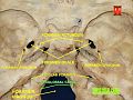
Hypoglossal canal
Base of the skull. Upper surface.

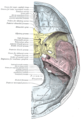
Median sagittal section through the occipital bone and first three cervical vertebræ.


See also[]
| Wikimedia Commons has media related to Hypoglossal canal. |
References[]
- ^ Takahashi, H.; Tanaka, H.; Fujita, N.; Tomiyama, N. (2014). "Bilateral persistent hypoglossal arteries: MRI findings". British Journal of Radiology. 85 (1010): e46-8. doi:10.1259/bjr/21939976. PMC 3473955. PMID 22308227.
- ^ Richards, Alan T. (2015). "14 - Surgical Exposures for the Nerves of the Neck". Nerves and Nerve Injuries. Vol. 2: Pain, Treatment, Injury, Disease and Future Directions. Academic Press. pp. 201–213. doi:10.1016/B978-0-12-802653-3.00063-4. ISBN 978-0-12-802653-3.
- ^ Voyvodic, F; Whyte, A.; Slavotinek, J. (1995). "The hypoglossal canal: normal MR enhancement pattern". American Journal of Neuroradiology. 16 (8): 1707–1710. PMID 7502978.
- ^ Katsuta, T.; Matsushima, T.; Wen, H.T.; Rhoton, A.L. (2000). "Trajectory of the hypoglossal nerve in the hypoglossal canal: significance for the transcondylar approach". Neurologia Medico-Chirurgica. 40 (4): 206–210. doi:10.2176/nmc.40.206. PMID 10853319.
- ^ 02.24.99 - Graduate Students Discredit Theory that Neanderthals Could Talk
External links[]
- Anatomy figure: 22:5b-15 at Human Anatomy Online, SUNY Downstate Medical Center
- cranialnerves at The Anatomy Lesson by Wesley Norman (Georgetown University) (XII)
- "Anatomy diagram: 34257.000-1". Roche Lexicon - illustrated navigator. Elsevier. Archived from the original on 2012-07-22.
- "Anatomy diagram: 34257.000-2". Roche Lexicon - illustrated navigator. Elsevier. Archived from the original on 2013-06-22.
- Image at uwo.ca
| Authority control: Scientific databases |
|---|
- Foramina of the skull