Foramen magnum
This article includes a list of general references, but it remains largely unverified because it lacks sufficient corresponding inline citations. (January 2013) |
| Foramen magnum | |
|---|---|
 Upper surface of base of the skull. The hole indicated by an arrow is the foramen magnum | |
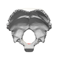
 Occipital bone. Inner surface. | |
| Details | |
| Part of | occipital bone |
| System | skeletal |
| Identifiers | |
| Latin | Foramen magnum |
| MeSH | D005539 |
| TA98 | A02.1.04.002 |
| TA2 | 553 |
| FMA | 75306 |
| Anatomical terms of bone | |
The foramen magnum (Latin: great hole) is a large, oval-shaped opening in the occipital bone of the skull. It is one of the several oval or circular openings (foramina) in the base of the skull. The spinal cord, an extension of the medulla oblongata, passes through the foramen magnum as it exits the cranial cavity. Apart from the transmission of the medulla oblongata and its membranes, the foramen magnum transmits the vertebral arteries, the anterior and posterior spinal arteries, the tectorial membranes and alar ligaments. It also transmits the accessory nerve into the skull.
The foramen magnum is a very important feature in bipedal mammals. One of the attributes of a biped's foramen magnum is a forward shift of the anterior border of the cerebellar tentorium; this is caused by the shortening of the cranial base. Studies on the foramen magnum position have shown a connection to the functional influences of both posture and locomotion. The forward shift of the foramen magnum is apparent in bipedal hominins→, including modern humans, Australopithecus africanus, and Paranthropus boisei. This common feature of bipedal hominins is the driving argument used by Michel Brunet that Sahelanthropus tchadensis was also bipedal, and may be the earliest known bipedal ape. The discovery of this feature has given scientists another form of identifying bipedal mammals.[1]
Structure[]
The foramen magnum is a large, oval-shaped opening (foramen) in the occipital bone of the skull.[2] It is present in humans, and in many other animals. Anteriorly, it is bounded by the basiocciput.[2] Posteriorly, it is bounded by the supraocciput.[2] Laterally, it is bounded by the occipital condyles.[2]
On the occipital bone, the foramen magnum presents two midline cephalometric landmarks. The opisthion is the midpoint on the posterior margin of the foramen magnum. The basion is located at the midpoint on the anterior margin of the foramen magnum.
The alar ligament, which is attached on each side to the tubercle of occipital condyle, divides the foramen magnum into an anterior smaller compartment and a posterior larger compartment.[3]
Variation[]
The foramen magnum varies in size between individuals. Earlier ossification of the occipital bone leads to a smaller foramen.[2]
Function[]
The foramen magnum transmits a number of important structures between the neck and the neurocranium.
Structures passing through anterior compartment (osseoligamentous compartment) include:
- the apical ligament and tip of the dens.
- the upper band of cruciate ligament of the atlas (C1 vertebra).
- the membrana tectoria.
Structures passing through posterior compartment (neurovascular compartment) include:
- the lower end of medulla oblongata, surrounded by meninges.
- the fourth part of the vertebral artery, surrounded by sympathetic plexus of nerves.
- accessory nerves.
- anterior and posterior spinal arteries.
- the tonsil of the cerebellum (occasionally) as in a tonsillar herniation known as a Chiari malformation.
Clinical significance[]
The foramen magnum may be too large, too small, or the wrong shape.[2] A small foramen magnum can cause neurological problems, and the reduced circulation of cerebrospinal fluid can cause hydrocephalus.[2] This may be treated with suboccipital craniectomy.[2]
Other animals[]
In humans, the foramen magnum is farther underneath the head than in the other great apes. Thus, in humans, the neck muscles (including the occipitofrontalis muscle) do not need to be as robust in order to hold the head upright. Comparisons of the position of the foramen magnum in early hominid species are useful to determine how comfortable a particular species was when walking on two limbs (bipedalism) rather than four (quadrupedalism).
The jerboa, a bipedal rodent, also has a foramen magnum.[4]
The foramen magnum varies in size and shape when comparing different populations to each other. In humans, men tend to have a larger sized foramen magnum than women, but the overall shape is consistent.[5]
Additional images[]

Skull seen from below. The hole through which the medulla (shown in red) is passing is foramen magnum.

Opisthion shown in red
Occipital bone inner surface (basion shown in red)


See also[]
References[]
![]() This article incorporates text in the public domain from page 129 of the 20th edition of Gray's Anatomy (1918)
This article incorporates text in the public domain from page 129 of the 20th edition of Gray's Anatomy (1918)
- ^ Russo, Gabrielle A.; Kirk, Christopher E. (November 2013). "Foramen magnum position in bipedal mammals". Journal of Human Evolution. 65 (5): 656–670. CiteSeerX 10.1.1.591.2458. doi:10.1016/j.jhevol.2013.07.007. PMID 24055116.
- ^ a b c d e f g h Sanchez, Pedro; Graham, John M. (2017). "31 - Congenital Anomalies of the Skull". Swaiman's Pediatric Neurology (6th ed.). Elsevier. pp. 233–241. doi:10.1016/B978-0-323-37101-8.00031-X. ISBN 978-0-323-37101-8.
- ^ Dutta, Asim Kumar (2013). Essentials of Human Anatomy Head & Neck. kolkata: Current books international. pp. 56–57. ISBN 978-81-86793-79-4.
- ^ Russo, Gabrielle A.; Kirk, E. Christopher (2013). "Foramen magnum position in bipedal mammals". Journal of Human Evolution. 65 (5): 656–70. CiteSeerX 10.1.1.591.2458. doi:10.1016/j.jhevol.2013.07.007. PMID 24055116. Lay summary – Phys.org (September 27, 2013).
- ^ Zdilla, Matthew J; Russell, Michelle L; Bliss, Kaitlyn N; Mangus, Kelsey R; Koons, Aaron W (2017). "The size and shape of the foramen magnum in man". Journal of Craniovertebral Junction & Spine. 8 (3): 205–221. doi:10.4103/jcvjs.JCVJS_62_17. ISSN 0974-8237. PMC 5634107. PMID 29021672.
External links[]
| Wikimedia Commons has media related to Foramen magnum. |
- "Anatomy diagram: 34257.000-1". Roche Lexicon - illustrated navigator. Elsevier. Archived from the original on 2012-07-22.
- Diagram 1
- Diagram 2
- 3D animation showing position of basion on YouTube
Authority control | |
|---|---|
| Scientific databases | |
| Other | |
- Wikipedia articles incorporating text from the 20th edition of Gray's Anatomy (1918)
- Foramina of the skull