Fimbria (bacteriology)
It has been suggested that this article be merged into Pilus. (Discuss) Proposed since December 2020. |

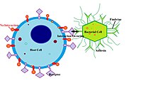
In bacteriology, a fimbria (Latin for 'fringe', plural fimbriae), also referred to as an "attachment pilus" by some scientists, is a short appendage found on many Gram-negative and some Gram-positive bacteria, and that is thinner and shorter than a flagellum. This appendage ranges from 3–10 nanometers in diameter and can be as much as several micrometers long. Fimbriae are used by bacteria to adhere to one another and to adhere to animal cells and some inanimate objects. A bacterium can have as many as 1,000 fimbriae. Fimbriae are only visible with the use of an electron microscope. They may be straight or flexible.
Fimbriae possess adhesins which attach them to some sort of substratum so that the bacteria can withstand shear forces and obtain nutrients. For example, E. coli uses them to attach to mannose receptors.
Some aerobic bacteria form a very thin layer at the surface of a broth culture. This layer, called a pellicle, consists of many aerobic bacteria that adhere to the surface by their fimbriae. Thus, fimbriae allow the aerobic bacteria to remain both on the broth, from which they take nutrients, and near the air.
All fimbriae are pili; they are only called fimbriae because of their purpose.[1] The term "fimbria" can refer to many different (structural) types of pilus, as many different types of pili have been used for adhesion, a case of convergent evolution.[2] The Gene Ontology system does not treat fimbriae as a distinct type of appendage, using the generic pilus (GO:0009289) type instead.
Examples[]
"Gram-negative bacteria assemble functional amyloid surface fibers called curli."[4] Curli are a type of fimbriae.[5] Curli are composed of proteins called curlins.[4] Some of the genes involved are CsgA, CsgB, CsgC, CsgD, CsgE, CsgF, and CsgG.[4]
Another type are called type 1 fimbriae.[5] They contain FimH adhesins at the "tips". The chaperone-usher pathway is responsible for moving many types of fimbriae out of the cell, including type 1 fimbriae[6] and the P fimbriae.[7]
Virulence[]
Fimbriae are one of the primary mechanisms of virulence for E. coli, Bordetella pertussis, Staphylococcus and Streptococcus bacteria. Their presence greatly enhances the bacteria's ability to attach to the host and cause disease.[8]
See also[]
References[]
- ^ Proft, T.; Baker, E. N. (February 2009). "Pili in Gram-negative and Gram-positive bacteria — structure, assembly and their role in disease". Cellular and Molecular Life Sciences. 66 (4): 613–635. doi:10.1007/s00018-008-8477-4. PMID 18953686. S2CID 860681.
- ^ Chagnot, C; Zorgani, MA; Astruc, T; Desvaux, M (14 October 2013). "Proteinaceous determinants of surface colonization in bacteria: bacterial adhesion and biofilm formation from a protein secretion perspective". Frontiers in Microbiology. 4: 303. doi:10.3389/fmicb.2013.00303. PMC 3796261. PMID 24133488.
- ^ WI, Kenneth Todar, Madison. "Colonization and Invasion by Bacterial Pathogens". www.textbookofbacteriology.net. Retrieved 2016-12-03.
- ^ a b c Epstein, EA; Reizian, MA; Chapman, MR (2009), "Spatial clustering of the curlin secretion lipoprotein requires curli fiber assembly.", J Bacteriol, 191 (2): 608–615, doi:10.1128/JB.01244-08, PMC 2620823, PMID 19011034.
- ^ a b Cookson, AL; Cooley, WA; Woodward, MJ (2002), "The role of type 1 and curli fimbriae of Shiga toxin-producing Escherichia coli in adherence to abiotic surfaces", Int J Med Microbiol, 292 (3–4): 195–205, doi:10.1078/1438-4221-00203, PMID 12398210.
- ^ Kolenda, Rafal; Ugorski, Maciej; Grzymajlo, Krzysztof (14 May 2019). "Everything You Always Wanted to Know About Salmonella Type 1 Fimbriae, but Were Afraid to Ask". Frontiers in Microbiology. 10: 1017. doi:10.3389/fmicb.2019.01017. PMC 6527747. PMID 31139165.
- ^ Rice JC, Peng T, Spence JS, Wang HQ, Goldblum RM, Corthésy B, Nowicki BJ (December 2005). "Pyelonephritic Escherichia coli expressing P fimbriae decrease immune response of the mouse kidney". Journal of the American Society of Nephrology. 16 (12): 3583–91. doi:10.1681/ASN.2005030243. PMID 16236807.
- ^ Connell I, Agace W, Klemm P, Schembri M, Mărild S, Svanborg C (September 1996). "Type 1 fimbrial expression enhances Escherichia coli virulence for the urinary tract". Proc. Natl. Acad. Sci. U.S.A. 93 (18): 9827–32. Bibcode:1996PNAS...93.9827C. doi:10.1073/pnas.93.18.9827. PMC 38514. PMID 8790416.
External links[]
- Fimbriae+Proteins at the US National Library of Medicine Medical Subject Headings (MeSH)
| Medical microbiology | |||||||
|---|---|---|---|---|---|---|---|
| Biochemistry and ecology |
| ||||||
| Shape | |||||||
| Structure |
| ||||||
| Taxonomy and evolution |
| ||||||
| |||||||
- Bacteriology