Alpha II-spectrin, also known as Spectrin alpha chain, brain is a protein that in humans is encoded by the SPTAN1gene.[5][6][7] Alpha II-spectrin is expressed in a variety of tissues, and is highly expressed in cardiac muscle at Z-disc structures, costameres and at the sarcolemma membrane. Mutations in alpha II-spectrin have been associated with early infantile epileptic encephalopathy-5, and alpha II-spectrin may be a valuable biomarker for Guillain–Barré syndrome and infantile congenital heart disease.
Alternate splicing of alpha II-spectrin has been documented and results in multiple transcript variants; specifically, cardiomyocytes have four identified alpha II-spectrin splice variants.[8][9] As opposed to alpha I-spectrin that is principally found in erythrocytes,[10] alpha II-spectrin is expressed in most tissues. In cardiac tissue, alpha II-spectrin is found in myocytes at Z-discs, costameres, and the sarcolemma membrane,[11][12][13] and in cardiac fibroblasts along the surface of the cytoskeletal network.[14] Alpha II-spectrin most commonly exists in a heterodimer with alpha II and beta II spectrin subunits; and dimers typically self-associate and heterotetramerize.[5][15][16]
Function[]
The spectrins are a family of widely distributed cytoskeletal proteins which are involved in actin crosslinking, cell adhesion, intercellular communication and cell cycle regulation.[17][18][19] Though a role in cardiac muscle is not well understood, it is likely that alpha II-spectrin is involved in organizing sub-sarcolemmal domains and stabilizing sarcolemmal membranes against the stresses associated with continuous cardiac contraction.[16] Functional diversity of alpha II-spectrin is manifest through its four splice variants. First, a cardiac-specific, 21 amino acid sequence insert in the 21st spectrin repeat, termed alpha II-cardi+, was identified as an insert that modulates affinity of alpha II-spectrin for binding beta-spectrins and regulates myocyte growth and differentiation.[8] Secondly, another insert of 20 amino acids in the 10th spectrin repeat, termed SH3i+, contains protein kinase A and protein kinase C phosphorylation sites and modulates Ca2+-dependent cleavage of spectrin and protein-protein interaction properties.[20] Thirdly, an insert of five amino acids in the fifteenth spectrin motif bears a highly antigenic epitope resembling an ankyrin-like p53 binding protein binding site.[8][21] Fourthly, a six amino acid insert in the twenty-first spectrin motif with unknown function has been reported.[11][22]
Alpha II-spectrin gene expression has been shown to be upregulated in cardiac fibroblasts in response to Angiotensin II-induced cardiac remodeling.[23]
In animal models of disease and injury, alpha II-spectrin has been implicated in diverse functions. In a canine model of hypothermic circulatory arrest, alpha II-spectrin breakdown products have shown to be relevant markers of neurologic injury post-cardiac surgery.[24]
Clinical significance[]
Mutations in SPTAN1 are the cause of early infantile epileptic encephalopathy-5.[25]
Alpha II-spectrin has shown promising utility as a biomarker for brain necrosis and apoptosis in infants with congenital heart disease; breakdown products of alpha II-spectrin have been detected in the serum of neonates in the perioperative period and following open-heart surgery.[26] Elevated protein expression of alpha II-spectrin has been detected in cerebrospinal fluid in patients with Guillain–Barré syndrome.[27]
^Bennett PM, Baines AJ, Lecomte MC, Maggs AM, Pinder JC (2004). "Not just a plasma membrane protein: in cardiac muscle cells alpha-II spectrin also shows a close association with myofibrils". Journal of Muscle Research and Cell Motility. 25 (2): 119–26. doi:10.1023/b:jure.0000035892.77399.51. PMID15360127. S2CID10297147.
^Sormunen R (Sep 1993). "Alpha-spectrin in detergent-extracted whole-mount cytoskeletons of chicken embryo heart fibroblasts". The Histochemical Journal. 25 (9): 678–86. doi:10.1007/bf00157882. PMID8226104. S2CID34132236.
^Sridharan DM, McMahon LW, Lambert MW (Nov 2006). "alphaII-Spectrin interacts with five groups of functionally important proteins in the nucleus". Cell Biology International. 30 (11): 866–78. doi:10.1016/j.cellbi.2006.06.005. PMID16889989. S2CID28863657.
^Wang XF, Gao GD, Liu J, Guo R, Lin YX, Chu YL, Han FC, Zhang WH, Bai YJ (2006). "Identification of differentially expressed genes induced by angiotensin II in rat cardiac fibroblasts". Clinical and Experimental Pharmacology & Physiology. 33 (1–2): 41–6. doi:10.1111/j.1440-1681.2006.04321.x. PMID16445697. S2CID21008341.
^Writzl K, Primec ZR, Stražišar BG, Osredkar D, Pečarič-Meglič N, Kranjc BS, Nishiyama K, Matsumoto N, Saitsu H (Jun 2012). "Early onset West syndrome with severe hypomyelination and coloboma-like optic discs in a girl with SPTAN1 mutation". Epilepsia. 53 (6): e106–10. doi:10.1111/j.1528-1167.2012.03437.x. PMID22429196. S2CID20216273.
^Hirai H, Matsuda S (September 1999). "Interaction of the C-terminal domain of delta glutamate receptor with spectrin in the dendritic spines of cultured Purkinje cells". Neurosci. Res. 34 (4): 281–7. doi:10.1016/s0168-0102(99)00061-9. PMID10576550. S2CID45794233.
Chow CW (1999). "Regulation and intracellular localization of the epithelial isoforms of the Na+/H+ exchangers NHE2 and NHE3". Clinical and Investigative Medicine. 22 (5): 195–206. PMID10579058.
Hayashi Y, Arakaki R, Ishimaru N (2003). "The role of caspase cascade on the development of primary Sjögren's syndrome". J. Med. Invest. 50 (1–2): 32–8. PMID12630566.
Bennett V (1979). "Immunoreactive forms of human erythrocyte ankyrin are present in diverse cells and tissues". Nature. 281 (5732): 597–9. Bibcode:1979Natur.281..597B. doi:10.1038/281597a0. PMID492324. S2CID263106.
Langley RC, Cohen CM (1986). "Association of spectrin with desmin intermediate filaments". J. Cell. Biochem. 30 (2): 101–9. doi:10.1002/jcb.240300202. PMID2939097. S2CID25080821.
Cianci CD, Giorgi M, Morrow JS (1988). "Phosphorylation of ankyrin down-regulates its cooperative interaction with spectrin and protein 3". J. Cell. Biochem. 37 (3): 301–15. doi:10.1002/jcb.240370305. PMID2970468. S2CID42349239.
McMahon AP, Giebelhaus DH, Champion JE, Bailes JA, Lacey S, Carritt B, Henchman SK, Moon RT (1987). "cDNA cloning, sequencing and chromosome mapping of a non-erythroid spectrin, human alpha-fodrin". Differentiation. 34 (1): 68–78. doi:10.1111/j.1432-0436.1987.tb00052.x. PMID3038643.
Frappier T, Regnouf F, Pradel LA (1988). "Binding of brain spectrin to the 70-kDa neurofilament subunit protein". Eur. J. Biochem. 169 (3): 651–7. doi:10.1111/j.1432-1033.1987.tb13657.x. PMID3121319.
McMahon AP, Moon RT (1988). "Structure and evolution of a non-erythroid spectrin, human alpha-fodrin". Biochem. Soc. Trans. 15 (5): 804–7. doi:10.1042/bst0150804. PMID3691949.
Lundberg S, Björk J, Löfvenberg L, Backman L (1995). "Cloning, expression and characterization of two putative calcium-binding sites in human non-erythroid alpha-spectrin". Eur. J. Biochem. 230 (2): 658–65. doi:10.1111/j.1432-1033.1995.0658h.x. PMID7607240.
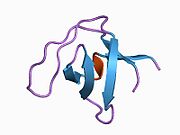
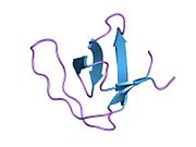
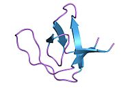
1m8m: SOLID-STATE MAS NMR STRUCTURE OF THE A-SPECTRIN SH3 DOMAIN
1pwt: THERMODYNAMIC ANALYSIS OF ALPHA-SPECTRIN SH3 AND TWO OF ITS CIRCULAR PERMUTANTS WITH DIFFERENT LOOP LENGTHS: DISCERNING THE REASONS FOR RAPID FOLDING IN PROTEINS
1qkw: ALPHA-SPECTRIN SRC HOMOLOGY 3 DOMAIN, N47G MUTANT IN THE DISTAL LOOP.
1qkx: ALPHA-SPECTRIN SRC HOMOLOGY 3 DOMAIN, N47A MUTANT IN THE DISTAL LOOP.
1shg: CRYSTAL STRUCTURE OF A SRC-HOMOLOGY 3 (SH3) DOMAIN
1u06: crystal structure of chicken alpha-spectrin SH3 domain
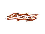
1u4q: Crystal Structure of Repeats 15, 16 and 17 of Chicken Brain Alpha Spectrin
1u5p: Crystal Structure of Repeats 15 and 16 of Chicken Brain Alpha Spectrin
1uue: A-SPECTRIN SH3 DOMAIN (V44T, D48G MUTANT)
2cdt: ALPHA-SPECTRIN SH3 DOMAIN A56S MUTANT
2f2v: alpha-spectrin SH3 domain A56G mutant
2f2w: alpha-spectrin SH3 domain R21A mutant
2f2x: alpha-spectrin SH3 domain R21G mutant
2fot: Crystal structure of the complex between calmodulin and alphaII-spectrin
2jm8: R21A Spc-SH3 free
2jm9: R21A Spc-SH3 bound
2jma: R21A Spc-SH3:P41 complex
2nuz: crystal structure of alpha spectrin SH3 domain measured at room temperature