Squalene-hopene cyclase
| Squalene-hopene cyclase | |||||||||
|---|---|---|---|---|---|---|---|---|---|
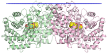 The crystallographic structure of the squalene-hopene cyclase dimer, with the membrane position indicated in blue, the two monomers in green and pink, and a substrate mimetic in the central cavity in yellow.[1] | |||||||||
| Identifiers | |||||||||
| EC no. | 5.4.99.17 | ||||||||
| CAS no. | 76600-69-6 | ||||||||
| Databases | |||||||||
| IntEnz | IntEnz view | ||||||||
| BRENDA | BRENDA entry | ||||||||
| ExPASy | NiceZyme view | ||||||||
| KEGG | KEGG entry | ||||||||
| MetaCyc | metabolic pathway | ||||||||
| PRIAM | profile | ||||||||
| PDB structures | RCSB PDB PDBe PDBsum | ||||||||
| |||||||||
Squalene-hopene cyclase (SHC) (EC 5.4.99.17) or hopan-22-ol hydro-lyase is a prokaryotic enzyme in the terpene cyclase/mutase family. It catalyzes the interconversion of an acyclic squalene molecule into a pentacyclic triterpenes hopene and hopanol in the ratio of 5:1.[2][3][4][5][6] This enzyme catalyses the following chemical reactions.
- squalene hop-22(29)-ene

- squalene + H2O hopan-22-ol
Squalene-hopene cyclase is important because its products, the hopenoids, are very much like sterols in eukaryotes in that they condense lipid membranes and reduce permeability. In the case of prokaryotes, they provide stability in the face of high temperatures and extreme acidity due to the rigid ring structures.[7] Indeed, upregulation of squalene-hopene cyclase occurs in certain bacteria in the presence of hot or acidic environments.[8][9]

Introduction[]
Squalene-hopene cyclase is found in a large number of bacteria but is most readily isolated from the thermophilic bacterium Alicyclobacillus acidocaldarius.[10] It catalyses the conversion of the acyclic molecule of squalene into the pentacyclic triterpenes of hopene and hopanol.

Squalene-hopene cyclase is thought to be an evolutionary progenitor of many classes of eukaryotic and prokaryotic sterol cyclases.[6] Oxidosqualene cyclases, which are eukaryotic analogs of SHC require oxygen for their reaction demonstrating a much later evolution when the atmosphere began accumulating oxygen. Squalene-hopene cyclase functions in a hypoxic environment suggesting a much earlier presence.[11]
Structure[]
Squalene-hopene cyclase is a membrane-associated 70-75kDa protein composed of 631 amino acids and seven PTFB repeats. It exists as a monotopic homodimer.[1]
Mechanism[]
The formation of the hopene skeleton is one of the most complex single step reactions in biochemistry.[12] In a single step, 13 covalent bonds are broken or formed, 9 chiral centers are established, and 5 rings are produced.[13] Squalene–hopene cyclase catalyses the conversion of the acyclic molecule of squalene into the pentacyclic triterpenes of hopene and hopanol. These products appear in the ratio 5:1. Hopene synthesis begins with binding squalene in an all pre-chair conformation and is followed by the formation of five C-C bonds.[14] These sequential ring-forming reaction steps are initiated by an electrophilic attack of an acidic proton on one of the two terminal double bonds. The polycyclic formation is completed when a proton is eliminated from the alternative terminal methyl group of squalene via acceptance by a water molecule.[5] This base is known as the front water. Other water molecules work to enhance polarization (back waters) and construct hydrogen bonds between seven residues—T41, E45, E93, R127, Q262, W133 and Y267. Front water also plays a role in determining the end product. If it stores the proton generated from either Methyl group 29 or 30 to form hopene. However, hopanol is produced in lesser quantities if instead of accepting the proton, water contributes a hydroxyl to the C-22 cation of the A-ring.[15]

During the formation of rings A through D, there is very little conformational change. The reaction therefore requires no intermediate and can take place in one step. However, ring E formation is hindered by an entropic barrier, which may explain its absence in the tetracyclic steroids.[5]
Active site[]
The SHC active site is located in a central cavity within the region of the protein adjacent to the membrane, and is accessed by the substrate via a non-polar channel.[16] The active site is notably surrounded by aromatic residues forming a cavity that comfortably fits the squalene molecule when folded into a productive conformation. The catalytic mechanism uses coupled aspartate and histidine residues to initiate the cyclization reaction by protonating at C3 and deprotonating at C29, proceeding through a discrete series of carbocation intermediates.[1][17] The enzyme can be inactivated by mutation of catalytic aspartates.[18]
Thermodynamics[]
This enzyme is unusually exothermic with an energy release of 40-50kcal/mol, well beyond the protein stabilization energy. This is thought to melt a lipid side channel through which the bulky product exits. In order to maintain its structural integrity, some scientists believe that the enzyme’s 7-8 non-tandem repeat QW motifs (Q is glutamine and W is tryptophan) that connect numerous surface α helices tighten the protein structure and prevent denaturing.[1]

References[]
- ^ a b c d Wendt KU, Poralla K, Schulz GE (September 1997). "Structure and function of a squalene cyclase". Science. 277 (5333): 1811–5. doi:10.1126/science.277.5333.1811. PMID 9295270.
- ^ Hoshino T, Sato T (February 2002). "Squalene-hopene cyclase: catalytic mechanism and substrate recognition". Chemical Communications (4): 291–301. doi:10.1039/b108995c. PMID 12120044.
- ^ Hoshino T, Nakano S, Kondo T, Sato T, Miyoshi A (May 2004). "Squalene-hopene cyclase: final deprotonation reaction, conformational analysis for the cyclization of (3R,S)-2,3-oxidosqualene and further evidence for the requirement of an isopropylidene moiety both for initiation of the polycyclization cascade and for the formation of the 5-membered E-ring". Organic & Biomolecular Chemistry. 2 (10): 1456–70. doi:10.1039/b401172d. PMID 15136801.
- ^ Sato T, Kouda M, Hoshino T (March 2004). "Site-directed mutagenesis experiments on the putative deprotonation site of squalene-hopene cyclase from Alicyclobacillus acidocaldarius". Bioscience, Biotechnology, and Biochemistry. 68 (3): 728–38. doi:10.1271/bbb.68.728. PMID 15056909.
- ^ a b c Reinert DJ, Balliano G, Schulz GE (January 2004). "Conversion of squalene to the pentacarbocyclic hopene". Chemistry & Biology. 11 (1): 121–6. doi:10.1016/j.chembiol.2003.12.013. PMID 15113001.
- ^ a b Pearson A, Budin M, Brocks JJ (December 2003). "Phylogenetic and biochemical evidence for sterol synthesis in the bacterium Gemmata obscuriglobus". Proceedings of the National Academy of Sciences of the United States of America. 100 (26): 15352–7. Bibcode:2003PNAS..10015352P. doi:10.1073/pnas.2536559100. PMC 307571. PMID 14660793.
- ^ Kannenberg, E.; Poralla, K. (1999). "Hopanoid biosynthesis and function in bacteria". Naturwissenschaften. 86 (4): 168–176. Bibcode:1999NW.....86..168K. doi:10.1007/s001140050592. S2CID 21596134.
- ^ Ourisson G, Rohmer M, Poralla K (1987). "Prokaryotic hopanoids and other polyterpenoid sterol surrogates". Annual Review of Microbiology. 41: 301–33. doi:10.1146/annurev.mi.41.100187.001505. PMID 3120639.
- ^ Sahm H, Rohmer M, Bringer-Meyer S, Sprenger GA, Welle R (1993). "Biochemistry and physiology of hopanoids in bacteria". Advances in Microbial Physiology. 35: 247–73. doi:10.1016/s0065-2911(08)60100-9. ISBN 9780120277353. PMID 8310881.
- ^ Seckler, B.; Poralla, K. (1986). "Characterization and partial purification of squalene-hopene cyclase from Bacillus acidocaldarius". Biochimica et Biophysica Acta (BBA) - General Subjects. 881 (3): 356–363. doi:10.1016/0304-4165(86)90027-9.
- ^ Rohmer, M.; Bouvier, G.; Ourisson, G. (1979). "Molecular evolution of biomembranes: structural equivalents and phylogenetic precursors of sterols". Proceedings of the National Academy of Sciences. 76 (2): 847–851. Bibcode:1979PNAS...76..847R. doi:10.1073/pnas.76.2.847. PMC 383070. PMID 284408.
- ^ Siedenburg G, Jendrossek D (June 2011). "Squalene-hopene cyclases". Applied and Environmental Microbiology. 77 (12): 3905–15. Bibcode:2011ApEnM..77.3905S. doi:10.1128/aem.00300-11. PMC 3131620. PMID 21531832.
- ^ Corey EJ, Matsuda SP, Bartel B (December 1993). "Isolation of an Arabidopsis thaliana gene encoding cycloartenol synthase by functional expression in a yeast mutant lacking lanosterol synthase by the use of a chromatographic screen". Proceedings of the National Academy of Sciences of the United States of America. 90 (24): 11628–32. Bibcode:1993PNAS...9011628C. doi:10.1073/pnas.90.24.11628. PMC 48037. PMID 7505443.
- ^ Zheng YF, Abe I, Prestwich GD (April 1998). "Inhibition kinetics and affinity labeling of bacterial squalene:hopene cyclase by thia-substituted analogues of 2, 3-oxidosqualene". Biochemistry. 37 (17): 5981–7. doi:10.1021/bi9727343. PMID 9558334.
- ^ "BRENDA - Information on EC 4.2.1.129 - squalene-hopanol cyclase".
- ^ Gao Y, Honzatko RB, Peters RJ (October 2012). "Terpenoid synthase structures: a so far incomplete view of complex catalysis". Natural Product Reports. 29 (10): 1153–75. doi:10.1039/C2NP20059G. PMC 3448952. PMID 22907771.
- ^ Hoshino T, Sato T (February 2002). "Squalene-hopene cyclase: catalytic mechanism and substrate recognition". Chemical Communications (4): 291–301. doi:10.1039/B108995C. PMID 12120044.
- ^ Feil, C.; Sussmuth, R.; Jung, G.; et al. (1996). "Site-directed mutagenesis of putative active-site residues in squalene-hopene cyclase". European Journal of Biochemistry. 242 (1): 51–55. doi:10.1111/j.1432-1033.1996.0051r.x. PMID 8954152.
External links[]
- Squalene-hopene+cyclase at the US National Library of Medicine Medical Subject Headings (MeSH)
- EC 5.4.99